
Cell Preparation Microbial Contamination Control
- Addtime: 2025-08-01 / View: 273
Current Status of Cell Preparation Contamination
Cell culture technology is widely used in scientific research, teaching, clinical trials, and the production of cell therapy products. In 2025, the National Medical Products Administration's Food and Drug Inspection Center issued the "Guidelines for Production Inspection of Cell Therapy Products." Large-scale cell culture in the production of cell therapy products places extremely high demands on aseptic techniques and technical expertise. According to a survey conducted by the Mycoplasma Key Laboratory of the Jiangsu Veterinary Research Institute, the mycoplasma contamination rate in cell cultures ranges from 15% to 35%, with some reports suggesting rates as high as 65% to 80%.
Microbial contamination, including bacterial, fungal, and viral, and mycoplasma contamination, is a common risk in large-scale cell culture operations.
Bacterial contamination is the most common type of microbial contamination in cell culture. Bacteria are tiny, simple-looking, single-celled microorganisms belonging to the prokaryotic kingdom. They typically originate from the cell culture environment and rarely from contaminated culture fluids and serum. Bacteria grow and reproduce rapidly, sometimes even within a single generation. During their growth and reproduction, bacteria not only consume large quantities of nutrients but also produce numerous toxins. Nutrient depletion and toxin attacks are major causes of cell death in contaminated cells. Fungal contamination: Fungi are eukaryotic microorganisms dozens of times larger than bacteria. They are highly resistant to desiccation, sunlight, ultraviolet light, and some disinfectants. Contamination of yeast cultures is similar to bacterial contamination. Round or ovoid yeasts can be observed under an inverted phase-contrast microscope, and a fermentation-like odor similar to wine or sour can be detected upon opening the culture flask. Contamination of mold cultures can be observed under an inverted phase-contrast microscope, with hyphae growing like branches. When lush, they are visible to the naked eye and resemble clumps of cotton wool.
Viral contamination: Viruses are microorganisms that can pass through bacterial filters, are measured in nanometers in size, and cannot independently utilize external nutrients for self-reproduction. Viral contamination in cell culture can arise from latent infection in the original host cells or from negligence during culture operations, such as using unsterilized flasks.
Mycoplasma contamination: Characteristics of mycoplasma. Mycoplasmas, also known as mycoplasmas, are microorganisms intermediate in size between bacteria and viruses, with a diameter of approximately 0.2 μm. They lack a cell wall, except for a ring-shaped molecule and ribosomes. Mycoplasma membranes are highly flexible and highly resistant to physical factors such as pressure, osmotic pressure, and dehydration. They are highly heterogeneous, and a small fraction can pass through conventional sterilizing filters. These characteristics make mycoplasmas the most common and challenging contaminants in cell culture.
Fantong Cell Contamination Solutions
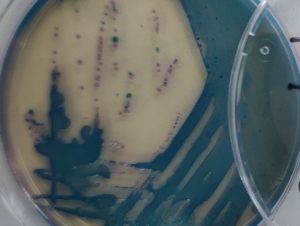
Contamination Source Tracing: Fantong engineers assist clients in detecting and identifying the microbial species that contaminate their products. They identify the microbial species and source through sterile sampling, isolation and culture, microscopic observation of morphology, and comparison of 16S/ITS gene sequencing with a database. The source table is then used to analyze the potential source.
Solution Development: Fantong's proprietary GHP disinfection technology effectively kills bacteria, spores, mycoplasmas, and mold spores without damaging process equipment.